3D spheroids
Moving beyond 2D: the use of 3D spheroids in drug discovery
What are 3D spheroids?
Cell-based assays are a staple technique of modern drug development, providing a controlled environment to explore disease biology and screen new compounds. Two-dimensional (2D) monolayers have been in use for many years, but their limitations are well recognised, particularly when it comes to modelling the architectures and microenvironments of solid tumours, or predicting in vivo drug responses. This has driven demand for more physiologically relevant in vitro models, with three-dimensional (3D) spheroids – tightly packed clusters of cells that grow in a spherical shape – emerging as a robust and scalable solution. Spheroids are already proving valuable across a range of applications, from oncology drug screening to safety pharmacology.
The limitations of traditional 2D cell cultures
2D cell culture models are well-established in drug discovery, but they lack the structural complexity of real tissues, meaning that the cells cannot interact with each other or their surroundings in a natural way. This is especially problematic when studying diseases such as cancers, where the tumour microenvironment plays a key role in how cells grow, spread and respond to treatment. As a result, drugs that appear promising in 2D systems often fail to perform in animal studies or clinical trials. The flat, uniform nature of 2D cultures simply doesn’t capture the biological conditions that influence how a drug is absorbed, distributed or resisted by the cells. This limits their predictive power, and can lead to costly setbacks later in the development process.
Why 3D spheroids offer a more powerful platform for drug discovery screening
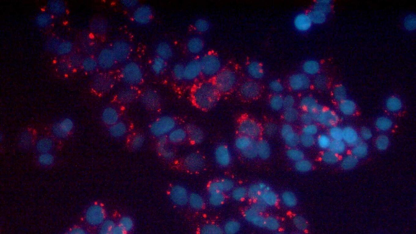
3D spheroids are tightly packed cell aggregates that grow in a spherical structure, either on their own (scaffold-free) or supported by a matrix (scaffold-based). This 3D organisation more closely mimics the structure and function of real tissues compared to flat 2D cultures, allowing for more natural cell-cell and cell-matrix interactions. Crucially, spheroids recreate important features of solid tumours, such as the formation of oxygen and nutrient gradients. As they grow, the outer layers remain well-nourished and proliferative, while the centre becomes more hypoxic and necrotic – just like in vivo tumours. This makes them especially valuable for modelling tumour behaviour and testing how drugs penetrate, diffuse and act within different zones of a tumour mass.
This increased physiological relevance means that 3D spheroids offer enhanced predictive power in drug screening applications. They provide a more accurate picture of how candidate compounds will behave in the body, including their ability to infiltrate tumour tissue, inhibit growth and prevent metastasis. Spheroids are also well suited to high-throughput screening, with 3D protocols increasingly adapted for automation and standardisation. This helps to identify more promising drug candidates earlier in development, reducing the number of false positives and costly failures in later-stage testing. Importantly, the improved data quality that spheroids provide can also reduce the need for animal testing in early-stage research, aligning with efforts to make drug development more ethical and efficient.
Looking ahead, 3D spheroids are also expected to play a growing role in personalised medicine. With advances in patient-derived cell cultures, it’s becoming increasingly feasible to generate spheroids from individual tumours, enabling researchers to test drug responses in vitro to guide more tailored treatment decisions. This could open up new possibilities for precision oncology and other targeted therapeutic areas.
Spheroids vs organoids vs organ-on-a-chip

The terminology around 3D cell culture models can be confusing, with spheroids, organoids and organ-on-a-chip sometimes used interchangeably. However, they represent distinct approaches, each with its own advantages and limitations:
- Spheroids are tightly packed clusters of cells, usually of a single cell type, that replicate a tumour or other in vivo microenvironment, allowing investigation of cell-cell, cell-matrix and cel-drug interactions. They offer the right balance of biological relevance, reproducibility and scalability for the majority of drug discovery applications, especially high-throughput screening.
- Organoids are miniaturised, simplified versions of organs, usually derived from stem cells. They self-organise into complex structures and contain multiple cell types, often mimicking organ function. Organoids are typically used for developmental biology or disease modelling, where multicellular complexity is essential, but usually lacks the scalability that is required for screening approaches.
- Organ-on-a-chip systems integrate living cells with microfluidic devices to simulate organ-level functions. These are more complex and expensive to use but are ideal for modelling physiological responses such as shear stress or drug metabolism across barriers.
Overcoming the challenges in spheroid production
Despite their many advantages, working with 3D spheroids can present several technical challenges, including:
- variability in size and shape, which can affect experimental reproducibility;
- inconsistent formation across different cell lines, requiring cell-specific optimisation;
- difficulty forming spheroids from small numbers of cells, especially with rare or patient-derived samples;
- challenges in creating multi-cell-type spheroids, such as co-cultures of tumour and stromal cells;
- and complexity in analysis, as their 3D structure can complicate imaging, staining and quantification.
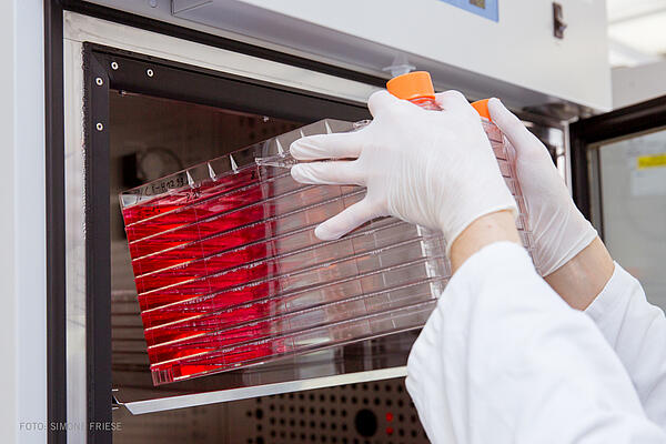
Standardisation of cell culture workflows is essential to address these challenges. Consistent protocols for cell seeding, media composition and culture conditions help to reduce variability, while consistent cell handling minimises batch-to-batch differences. The chosen method for spheroid formation – whether low-attachment plates, hanging drops or bioreactors – also has a significant impact on reproducibility. In parallel, new techniques are emerging to support high-throughput spheroid production. These include automated platforms and amplification methods that generate large numbers of uniform spheroids, making it easier to integrate them into drug screening pipelines at scale.
Future trends in 3D spheroid technology
3D spheroids are set to become an even more powerful and practical model, bridging the gap between traditional in vitro assays and complex in vivo biology. As 3D models become more embedded in drug discovery workflows, the focus is shifting towards improving scalability, reproducibility and ease of use. There is a clear industry-wide push for standardised platforms that can reliably generate high-quality spheroids across different cell types and applications, especially in high-throughput screening environments. Companies like acCELLerate are helping to drive this progress by developing ready-to-use solutions that simplify spheroid formation. This includes the large-scale preparation of cryopreserved tumour cell lines, supplied in aliquots that are optimised to generate uniform spheroids once plated. These innovations reduce day-to-day variability and help ensure consistent results across labs and experiments, driving the development of new therapies.
Author

Dr. Oliver Wehmeier
- eMail: Contact me
- Linkedin: My LinkedIn